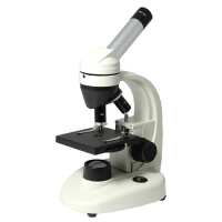
Instruction ManualBM0902 Biological Microscope
Instruction Manual-English.docQuick OverviewFinite. Total Magnification: 40-400X. 10X Eyepiece. 4X 10X 40X Achromatic Objective. Eye Tube Angle: 45°. Eyepiece Field of View:
Dia. 15mm. Illumination Type: LED Coaxial Transmitted Light. Input Voltage: AC 100-240V 50/60Hz. Suggested ApplicationsEducation & Student , Compound (High Power) , Elementary (K-8) , Kids, Forensics , Blood Analysis, Hair & Fiber AnalysisBM09020101 Monocular Biological MicroscopeOptical System SpecificationsOptical SystemFiniteMechanical Tube Length160mmSystem Optical Magnification40-400XTotal Magnification40-400XStandard Eyepiece10X EyepieceStandard Objective4X 10X 40X Achromatic ObjectiveSystem Field of View Dia. 0.375-3.75mmMonocular HeadEye Tube Optical SystemFiniteEye Tube TypeFor Compound MicroscopeEye Tube Angle45°Erect/Inverted ImageInverted ImageEye Tube Rotatable360° Degree RotatableEye Tube Inner Diameter Dia. 23.2mmEye Tube Fixing ModeWithout LockingEyepiece10X Eyepiece ( Dia. 23.2/FN15)Eyepiece TypeStandard EyepieceEyepiece Optical Magnification10XPlan EyepieceHuygens EyepieceEyepiece Size for Eye Tube Dia. 23.2mmEyepiece Field of View Dia. 15mmSurface TreatmentElectroplating BlackMaterialMetalColorBlackNet Weight0.02kg (0.04lbs)Biological Objective4X Achromatic ObjectiveObjective Optical SystemFiniteObjective Optical Magnification4XObjective TypeAchromatic ObjectiveObjective Parfocal Distance45mmObjective for Mechanical Tube Length160mmObjective Immersion MediaDry ObjectiveObjective Outer Diameter Dia. 17mmSurface TreatmentElectroplating BlackMaterialMetalColorBlackNet Weight0.01kg (0.02lbs)Applied FieldBM0902 Series Microscope10X Achromatic ObjectiveObjective Optical SystemFiniteObjective Optical Magnification10XObjective TypeAchromatic ObjectiveObjective Parfocal Distance45mmObjective for Mechanical Tube Length160mmObjective Immersion MediaDry ObjectiveObjective Outer Diameter Dia. 17mmSurface TreatmentElectroplating BlackMaterialMetalColorBlackNet Weight0.02kg (0.04lbs)Applied FieldBM0902 Series Microscope40X Achromatic ObjectiveObjective Optical SystemFiniteObjective Optical Magnification40XObjective TypeAchromatic ObjectiveObjective Parfocal Distance45mmObjective for Mechanical Tube Length160mmObjective Immersion MediaDry ObjectiveSpring Mounted ObjectiveSpring Mounted objectiveObjective Outer Diameter Dia. 17mmSurface TreatmentElectroplating BlackMaterialMetalColorBlackNet Weight0.01kg (0.02lbs)Applied FieldBM0902 Series MicroscopeNosepieceInward/Outward NosepieceNosepiece Inward Number of Holes on NosepieceTriple (3) HolesNosepiece Switch ModeManualNosepiece Screw Thread for Objective3/5 in. x1/40 in. Microscope StandStand Height195mmBase TypeIllumination BaseBase ShapeUnconventionalBase Dimensions165x135x30mmFocus ModeManualFocus Distance13mmCoarse Focus Distance per Rotation25mmFocusing Knob Tightness AdjustableTightness Not AdjustableMicroscope StageStage Platform Dimensions90x90mmStage Backlight Window Size Dia. 11mmNumber of Stage Clips1 PairMicroscope IlluminatorIllumination TypeLED Coaxial Transmitted LightTransmission LightBright FieldTransmission Light Source TypeLEDAperture Diaphragm Mounting PositionVertical IlluminatorField Diaphragm Mounting PositionVertical IlluminatorPower SupplyInput VoltageAC 100-240V 50/60HzOutput VoltageDC 5VPower Cord Connector TypeUSA 2 PinsPower Cable Length1.3mOther ParametersSurface TreatmentSpray PaintMaterialMetalColorWhiteNet Weight1.38kg (3.04lbs)Dimensions230x140x280mm (9.055x5.512x11.024 in. )SeriesBM0902BM09020101
.tb_part
{
width: 100%;
border: solid 0px #ccc;
font-size:12px;
}
.tb_part tr
{
height: 30px;
line-height: 30px;
}
.tb_part td
{
white-space: nowrap;
}
.tb_part_1 td
{
white-space: wrap;
}
.normal{
border-left:solid 2px black;
border-right:solid 2px black;
}
.tb_part tr.normal td
{
border:solid 1px #ccc;
padding-right:5px;
}
.tb_part tr.normal:nth-child(2){
border-top:solid 2px black;
}
.tb_part tr.normal:first-child{
border:none;
}
.tb_part tr.normal:first-child td{
border:none;
font-weight:bold;
font-size:16px;
}
.end-border{
border-bottom:solid 2px black;
}
.tb_part tr.memo,.tb_part tr.memo td{
border:none;
}
Technical InfoInstructionsBiological MicroscopeClose ΛBiological microscopes are compound microscopes that are primarily used to observe and study organisms and microorganisms. Biological microscopes were the earliest type of microscopes to be invented and the most widely used compound microscope today. Humans first used simple microscopes to observe tiny objects with a lens. Later, compound microscope were invented, which then used two lenses, i.e., one eyepiece and one objective lens for secondary imaging, to obtain a larger multiple of the image. Conventionally, we usually refer to microscopes that include various accessories such as phase contrast, fluorescence, and polarized light etc. as compound microscopes, to distinguish them from stereo microscopes. (Although stereo microscopes also have an eyepiece and an objective lens, they have two light paths, which presents a three-dimensional image).
The most basic biological microscope consists of an eyepiece, an objective lens, a microscope stage, and light source. Both the eyepiece and the objective lens are convex lenses. The objective lens first enlarges the object into a real image. The eyepiece then magnifies the real image again into a virtual image, and finally becomes an inverted magnified virtual image on the retina of the human eye. Biological microscopes are usually used to observe transparent or translucent objects, such as animal and plant cells, tissues, bacteria and microorganisms, as well as various kinds of tiny particles by means of sectioning. They are widely used in teaching, medicine, animal or plant research and industrial fields. Modern optical microscopes have made great progress in the wavelengths of various kinds of light; illumination forms, resolution, microscope functions, structure and comfort of image acquisition and analysis, and basically meet various research needs. According to the user's needs and the complexity of the product, general biological microscopes are divided into student-level, experimental-grade, and research-level biological microscopes.
Basic Structure of Biological Microscope A standard biological microscope usually has at least the following basic structures: 1. Objective lens - the closest imaging lens to the observed specimen. Objective lens determines the most important properties of the microscope imaging; such as wavelength and resolution of the object light. A microscope can have several objective lenses with different magnifications.
2. Eyepiece - the lens mounted on the upper end of the microscope tube; close to the observer's eyes. Generally, microscopes can have several eyepieces with different magnifications. 3. Light source - the light source of the biological microscope is under the microscope stage. According to different needs, a light source may include an illuminating light source (bulb), an aperture diaphragm, a condenser etc. The condenser is used to condense the illumination light and also increase the illumination brightness of the specimen. Aperture diaphragm, also called iris, is used to adjust the luminous flux of light. Under the aperture diaphragm, there is usually a circular filter holder, and the optical filters are placed according to needs. A simple microscope would not have an illuminating light source, it is illuminated by natural light, and a reflector is used to illuminate the object to be observed. 4. Microscope base - located at the bottom of the microscope; to support the lens body. Usually, the light source and the electrical appliances are installed inside the base and above the base. 5. Microscope body - used to connect and stand the various components of the entire microscope, and it is also the part the user holds when moving the microscope. 6. Microscope tube - an optical path channel connecting the eyepiece and the nosepiece of the microscope. 7. Nosepiece - the turntable under the microscope tube. The nosepiece usually has 3 to 4 circular holes for mounting objective lenses of different magnification; which can be rotated onto the optical axis of the microscope for use. 8. Microscope stage - where the specimen is placed for observation. There are usually two metal tablets on the mobile station, which are used to fix the specimen of the slide. There is also usually a pusher installed for moving the specimen. There are also microscope stages that can be moved directly in the XY direction. 9. Focus knob - used to adjust the distance between the objective lens and the microscope stage (sample) to bring the objective lens into focus to get a clear picture or image. The focus knob is usually mounted with the microscope stage to achieve the purpose of moving up and down focusing through the coarse focus knob and the fine focus knob.
Biological Microscope Quick Operation Steps Step 1. Install and Prepare: The configuration of the biological microscope is mostly standard. Carefully check the parts on the packing list and the information on the BoliOptics website to assemble and install the microscope.
The microscope should be placed on a solid and stable work surface with the tabletop kept steady, clean, and close to a power source. It is best to place the microscope out of direct sunlight. Generally speaking, the darker the environment, the better the image is observed by the microscope. Stray light greatly influences the imaging when the microscope is used for observation, as it can damage the specimen and can also accelerate the aging of the microscope surface and components.
Step 2. Turn on the light source: Connect the power source, turn on the power switch, and adjust the light source to a position where the brightness is moderate.
Step 3. Place the specimen (also known as the type or sample): Adjust the coarse focus knob first, and raise the objective lens to a higher position for easy placement of the specimen. Place the slide specimen of the observed object on the microscope stage. Note that the side of the cover slip is placed face up. Then use spring pressure to clamp on both ends of the slide to prevent the specimen from moving, and then adjust the knob through the XY direction of the microscope stage to move the general position of the part of the specimen to be observed to the center of the condenser.
Step 4. Adjust the parfocal of the high and low objective lens: First observe with low power objectives. Adjust the low power lens (such as 4X, 10X) from the objective lens or nosepiece to the optical axis. Then, adjust the focus knob to find the outline of the image. Because the low power objectives have a large field of view, it is easier to find the image and determine the part to be observed. At the same time, adjust the XY microscope stage hand button to find the position of the specimen to be observed. It should be noted that the image of the biological microscope in the field of view is usually an inverted image, that is, the specimen should be moved in the opposite direction when moving the specimen. Then, turn the nosepiece and gradually use the high power objective (such as 40X) to move to the observation position, and finally to the maximum magnification (such as 100X). During the process, continually adjust the fine adjustment knob to find the clearest image. With regard to the observation and use of the oil lens, it is generally carried out after the above steps, and finally make further accurate observation.
When changing from low power objectives to high power objectives, the object image can generally be seen, but it may not be very clear. When rotating to the maximum power objectives (such as 100X), only the fine focus knob should be used rather than the coarse focus knob, so as to avoid damage to the lens or the slide specimen. When the image of the maximum power objective is clear using a microscope with normal function, ensure that the low power objectives and the high power objectives are parfocal, and the focus knob is no longer adjusted. During operation, it is possible that the power of some of the objectives in the middle may not be parfocal. If so, you only need to adjust the fine focus knob slightly. Using a binocular microscope - If the observer's binocular vision is different, adjust it by the eyetube diopter of the eyepieces. Do not adjust the focus knob.
Step 5. Adjust the Light Source: Adjust the light intensity of the light source. Adjust the size of the diaphragm, the height of the condenser, the angle of the reflector. These adjustments need to be coordinated and adjusted with the power of objective in order to get a clear image. Under normal circumstances, the light of the stained specimen should be strong, and the light of the colorless or unstained specimen should be dim. When adjusting between high and low power objectives, the light for low power objectives for observation should be dim, and the light for high power objectives for observation should be strong.
Step 6. Replace the specimen: After observing the specimen - if you need to switch to another slide, you should first change the objectives back to low power, remove the slide before replacing it with a new one, and then adjust the focus again for observation. Do not change the specimen under the high power objectives as the working distance is very small, so as to prevent damage to the objective lens.
Step 7. Arranging the microscope after use: After observing with the microscope, the objective lenses should be moved away from the light-passing hole. Turn the nosepiece so that the V-shape between the lenses is slanted to both sides. Remove the sample. Check the light source of the microscope - adjust the aperture diaphragm to the maximum, adjust the brightness knob to the darkest, and then turn off the power button to prevent the instantaneous high current from burning out the light source when the power is turned on next time. Lower the microscope stage and check if any parts are damaged, if the objective lens is stained with water or oil, or if the objective body has stains or hand prints. Wipe the microscope clean, and check that the accessories are complete, the sample specimens are complete, and anything else is complete. After the final inspection is completed, cover the microscope with a dust cover or place the microscope into a box.
Biological microscopes are the basic structure of other forms of compound microscopes that are added with various kinds of accessories or attachments. Many principles and key points are fundamentally reflected in biological microscopes. FiniteClose ΛMicroscopes and components have two types of optical path design structures. One type is finite optical structural design, in which light passing through the objective lens is directed at the intermediate image plane (located in the front focal plane of the eyepiece) and converges at that point. The finite structure is an integrated design, with a compact structure, and it is a kind of economical microscope. Another type is infinite optical structural design, in which the light between the tube lens after passing the objective lens becomes "parallel light". Within this distance, various kinds of optical components necessary such as beam splitters or optical filters call be added, and at the same time, this kind of design has better imaging results. As the design is modular, it is also called modular microscope. The modular structure facilitates the addition of different imaging and lighting accessories in the middle of the system as required. The main components of infinite and finite, especially objective lens, are usually not interchangeable for use, and even if they can be imaged, the image quality will also have some defects.
The separative two-objective lens structure of the dual-light path of stereo microscope (SZ/FS microscope) is also known as Greenough. Parallel optical microscope uses a parallel structure (PZ microscope), which is different from the separative two-object lens structure, and because its objective lens is one and the same, it is therefore also known as the CMO common main objective. Mechanical Tube LengthClose ΛFor objective lens design of finite microscope, its mechanical tube length is the distance from the objective nosepiece shoulder of the objective lens to the eyepiece seat in the tubes, that is, the eyepiece shoulder.
There are two standards in the traditional microscope structure, namely, DIN and JIS. DIN (Deutsches Institute fur Normung) is a popular international standard for microscopes, using 195mm standard conjugate distance (also known as object to primary image distance, 36mm objective lens parfocal distance, and 146.5mm optical tube length.
JIS (Japanese Industrial Standard) is a standard adopted by some Japanese manufacturers, using 160mm standard conjugate distance (also known as object to primary image distance), 45mm objective lens parfocal distance), and 150mm optical tube length.
Using the same microscope standard design, the objective lenses can be used interchangeably. System Optical MagnificationClose ΛThe magnification of the objective lens refers to the lateral magnification, it is the ratio of the image to the real size after the original image is magnified by the instrument. This multiple refers to the length or width of the magnified object. System optical magnification is the product of the eyepiece and the objective lens (objective lens zoom set) of the optical imaging part within the system. Optical magnification = eyepiece multiple X objective lens/objective lens set
The maximum optical magnification of the microscope depends on the wavelength of the light to which the object is illuminated. The size of the object that can be observed must be greater than the wavelength of the light. Otherwise, the light cannot be reflected or transmitted, or recognized by the human eye. The shortest wavelength of ultraviolet light is 0.2 microns, so the resolution of the optical microscope in the visible range does not exceed 0.2 microns, or 200 nanometers. This size is converted to the magnification of the microscope, and it is the optical magnification of 2000X. Usually, the compound microscope can achieve 100X objective lens, the eyepiece is 20X, and the magnification can reach 2000X. If it is bigger, it will be called "invalid magnification", that is, the image is large, but the resolution is no longer increased, and no more details and information can be seen. Total MagnificationClose ΛTotal magnification is the magnification of the observed object finally obtained by the instrument. This magnification is often the product of the optical magnification and the electronic magnification. When it is only optically magnified, the total magnification will be the optical magnification.
Total magnification = optical magnification X electronic magnification Total magnification = (objective X photo eyepiece) X (display size / camera sensor target ) System Field of ViewClose ΛField of View, is also called FOV.
The field of view, or FOV, refers to the size of the object plane (i.e., the plane of the point of the observed object perpendicular to the optical axis), or of its conjugate plane (i.e., object to primary image distance), represented by a line value. System field of view is the size of the actual diameter of the image of the terminal display device of the instrument, such as the size of the image in the eyepiece or in the display.
Field of view number refers to the diameter of the field diaphragm of the objective lens, or the diameter of the image plane formed by the field diaphragm. Field of view number of objective lens = field of view number of eyepiece / (objective magnification / mechanical tube length)
Large field of view makes it easy to observe the full view and more range of the observed object, but the field of view (FOV) is inversely proportional to the magnification and inversely proportional to the resolution, that is, the larger the field of view, the smaller the magnification, and also the lower the resolution of the object to be observed. There are usually two ways to increase the field of view, one is to replace with an objective lens of a smaller multiple, or to replace with an eyepiece of a smaller multiple. Eye Tube AngleClose ΛUsually the Microscope Eyetube is 45°, some is 30°, Tiltable Eyetube Angle design of a microscope is also known as the ergonomics microscope. 0-30° or 0-45° is an ergonomic design. When the mechanical tube length / focal length of the tube of the microscope is relatively big, the microscope is relatively high, and the user's height or the seat of the work desk is not suitable, long-term use of microscope may cause sitting discomfort. Eyepiece tube with variable angle can freely adjust the angle without lowering the head. Especially when it is close to 0 degree and the human eye is close to horizontal viewing, long-time or long-term use can avoid fatigue damage to the cervical vertebra.Erect/Inverted ImageClose ΛAfter imaging through a set of objective lenses, the object observed and the image seen by the human eye is inverted. When the observed object is manipulated, move the specimen or object, the image will move in the opposite direction in the field of view. Most of the biological microscopes are reversed-phase designs. When needing to operate works with accurate direction, it is necessary to design it into a forward microscope. Generally stereo microscopes and metallurgical microscopes are all of erect image design. When observing through the camera and display, the erect and inverted image can be changed by the orientation of the camera. 360° Degree RotatableClose ΛThe eyepiece of the microscope can have different viewing or observing directions. When the position of the microscope is uncomfortable, the direction of the eyepiece tube of the microscope can be adjusted, to facilitate observation and operation.
Placement method of different viewing angles of the microscope: General direction: the support column is behind the object to be observed Reverse direction: the support column is in front of the object to be observed Lateral direction: the support column is on the side of the object to be observed Rotating eyepiece tube, different microscopes may have different methods, for some, the direction is confirmed when installing the eyepiece tube of the microscope, for some, by rotating the body of the microscope, and for some, by rotating the support member on the support or holder of the microscope.Eyepiece Optical MagnificationClose ΛEyepiece optical magnification is the visual magnification of the virtual image after initial imaging through the eyepiece. When the human eye observes through the eyepiece, the ratio of the tangent of the angle of view of the image and the tangent of the angle of view of the human eye when viewing or observing the object directly at the reference viewing distance is usually calculated according to 250 mm/focal length of eyepiece. The standard configuration of a general microscope is a 10X eyepiece. Usually, the magnification of the eyepiece of compound microscope is 5X, 8X, 10X, 12.5X, 16X, 20X. As stereo microscope has a low total magnification, its eyepiece magnification generally does not use 5X, but can achieve 25X, 30X and other much bigger magnification. Huygens EyepieceClose ΛThe Huygoens eyepiece is composed of two single-sided plano-convex lenses of the same type of glass, consisting of two convex lenses that have not been corrected by chromatic aberration; both are convex toward the objective lens, and the piece close to the eye is called eye piece, for magnification function; the other piece is called field lens, which makes the image brightness uniform. On the focal plane of the eye lens between the two lenses, there is a diaphragm, which is a parallax diaphragm, which also plays the role of the eyepiece to eliminate the stray light, and can also place the reticle or the pointer on this diaphragm.
Huygens eyepiece has no correction aberration. It is only suitable for use with low and medium achromatic objectives. They are simple in structure and low in cost, which is a commonly used eyepiece for low-end microscopes. Huygens eyepieces can be used for both observation and photography. When the image formed by the objective lens is within the focus of the eye lens, it becomes a magnified virtual image, microscopic observation can be performed; when the image formed by the objective lens is outside the focus of the eye lens, it becomes magnified real image, microscopic photography can be performed.Eyepiece Field of ViewClose ΛThe eyepiece field of view is the diameter of the field diaphragm of the eyepiece, or the diameter of the image plane of the field diaphragm imaged by the field diaphragm. The diameter of a large field of view can increase the viewing range, and see more detail in the field of view. However, if the field of view is too large, the spherical aberration and distortion around the eyepiece will increase, and the stray light around the field of view will affect the imaging effect. Objective Optical MagnificationClose ΛThe finite objective is the lateral magnification of the primary image formed by the objective at a prescribed distance.
Infinite objective is the lateral magnification of the real image produced by the combination of the objective and the tube lens.
Infinite objective magnification = tube lens focal length (mm) / objective focal length (mm)
Lateral magnification of the image, that is, the ratio of the size of the image to the size of the object. The larger the magnification of the objective, the higher the resolution, the smaller the corresponding field of view, and the shorter the working distance. Objective TypeClose ΛIn the case of polychromatic light imaging, the aberration caused by the light of different wavelengths becomes chromatic aberration. Achromatic aberration is to correct the axial chromatic aberration to the two line spectra (C line, F line); apochromatic aberration is to correct the three line spectra (C line, D line, F line). The objective is designed according to the achromaticity and the flatness of the field of view. It can be divided into the following categories.
Achromatic objective: achromatic objective has corrected the chromatic aberration, spherical aberration, and comatic aberration. The chromatic portion of the achromatic objective has corrected only red and green, so when using achromatic objective, yellow-green filters are often used to reduce aberrations. The aberration of the achromatic objective in the center of the field of view is basically corrected, and as its structure is simple, the cost is low, it is commonly used in a microscope.
Semi-plan achromatic objective: in addition to meeting the requirements of achromatic objective, the curvature of field and astigmatism of the objective should also be properly corrected. Plan achromatic objective: in addition to meeting the requirements of achromatic objectives, the curvature of field and astigmatism of the objective should also be well corrected. The plan objective provides a very good correction of the image plane curvature in the field of view of the objective, making the entire field of view smooth and easy to observe, especially in measurement it has achieved a more accurate effect.
Plan semi-apochromatic objective: in addition to meeting the requirements of plan achromatic objective, it is necessary to well correct the secondary spectrum of the objective (the axial chromatic aberration of the C line and the F line). Plan apochromatic objective: in addition to meeting the requirements of plan achromatic objective, it is necessary to very well correct the tertiary spectrum of the objective (the axial chromatic aberration of the C line, the D line and the F line) and spherochromatic aberration. The apochromatic aberration has corrected the chromatic aberration in the range of red, green and purple (basically the entire visible light), and there is basically no limitation on the imaging effect of the light source. Generally, the apochromatic aberration is used in a high magnification objective.
Objective Parfocal DistanceClose ΛObjective parfocal distance refers to the imaging distance between the objective shoulder and the uncovered object surface (referred to as the “object distance). It conforms to the microscope design, usually 45mm.
The objective of different magnifications of the compound microscope has different lengths; when the distance between the objective shoulder and the object distance is the same, the focal length may not be adjusted when converting to objectives of different magnifications.
Objective for Mechanical Tube LengthClose ΛObjective for mechanical tube length is a design parameter of the mechanical tube length of the microscope that the objective is suitable for.Objective Immersion MediaClose ΛThe use of different media between the objective and the object to be observed is to change and improve the resolution. For example, the refractive index of air is 1, water is 1.33, and cedar oil is 1.515. Therefore, when using an aqueous medium or cedar oil, a greater N.A. value can be obtained, thereby increasing the resolution of the objective. Air medium is called dry objective, where oil is used as medium iscalled oil immersion objective, and water medium is called water immersion objective. However, because of the working distance of the objective, when the working distance of the objective is too long, the use of liquid medium will be relatively more difficult, and it is generally used only on high magnification objective having a shorter working distance, such as objectives of 60X, 80X and 100X.
When using oil immersion objective, first add a drop of cedar oil (objective oil) on the cover glass, then adjust the focus (fine adjustment) knob, and carefully observe it from under the side of the objective of the microscope, until the oil immersion objective is immersed in the cedar oil and close to the cover glass of the specimen, then use the eyepiece to observe, and use the fine focus knob to lift the tube until the clear imageof the specimen is clearly seen. The cedar oil should be added in an appropriate amount. After the oil immersion objective is used, it is necessary to use a piece of lens wiping tissue to dip xylene to wipe off the cedar oil, and then wipe dry the lens thoroughly with a lens wiping tissue.
Spring Mounted ObjectiveClose ΛThe front end of the objective is equipped with a spring device. When the working distance of the objective is too short, focusing can easily make the objective contact the object to be observed, thereby damaging the object to be observed or the front lens. At this time, the spring acts to recover the front end of the objective lens. It is usually used on high magnification objectives with very short working distances.Illumination BaseClose ΛIllumination base is a modular light source component, suitable for microscope stand base that has no light source of itself, and it is usually dedicated components supporting some stands. Illumination base typically includes at least one bottom lighting, and there are also illumination base that includes the circuit portion of the upper light source. Focusing Knob Tightness AdjustableClose ΛDifferent microscope bodies, different human operations, and different requirements for observation and operation, all require adjustment of the pre-tightening force of the stand that support microscope body. Facing the stand just right, use both hands to reverse the force to adjust the tightness. (face the knob of one side just right, clockwise is to tighten, counterclockwise is to loosen) In general, after long-time use, the knob will be loose, and adjustment is necessary.
Stage Backlight Window SizeClose ΛStage backlight window size refers to the size of the window through which the transmitted light passes under the stage on the XY table plane of the stage. This window is usually covered with a piece of glass. For some stages with accuracy requirements in the XY horizontal direction, the horizontal plane of the glass can be adjusted by the height of the screws on the four corners below, and the consistency with the height of the stage plane is guaranteed. PackagingClose ΛAfter unpacking, carefully inspect the various random accessories and parts in the package to avoid omissions. In order to save space and ensure safety of components, some components will be placed outside the inner packaging box, so be careful of their inspection. For special packaging, it is generally after opening the box, all packaging boxes, protective foam, plastic bags should be kept for a period of time. If there is a problem during the return period, you can return or exchange the original. After the return period (usually 10-30 days, according to the manufacturer’s Instruction of Terms of Service), these packaging boxes may be disposed of if there is no problem.
Optical Data Microscope Optical Data SheetP/NObjectiveObjective Working DistanceEyepieceBM09022211 (10X Dia. 15mm)MagnificationField of View(mm)BM090232114X40X3.75mmBM0902331110X100X1.5mmBM0902351140X400X0.38mm1. Magnification=Objective Optical Magnification * Body Magnification * Eyepiece Optical Magnification2. Field of View=Eyepiece Field of View /Objective Optical Magnification*Body Magnification3. The Darker background items are Standard items, the white background items are
optional items.
About Boli Optics:
Boli Optics Microscope Supplier sells professional microscopes, microscope accessories, and magnifying lamps. We offer parts and accessories compatible with Leica, Olympus, Nikon, and Zeiss, and more. We supply research laboratories, medical centers, universities, industrial manufactures, factories, and more. Our engineers and technicians provide technical support and design & manufacture custom microscope products for your applications. Our products are manufactured under ISO 9001 quality control standards. We also provide OEM service. Since 1994, our talented team has been working in the optics industry and serving our customers whole-heartedly. Based in Southern California, we offer fast, same day shipping from our local warehouses.
Visit Product PageEmail:
sales@bolioptics.com
Location:
8762 Lanyard Court,
Rancho Cucamonga
California
91730