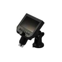
This Digital Microscope with LCD screen offers 1-600X magnification, with continuous smooth digital zoom in and out. It can capture images and record videos at 30 fps.3.6 megapixels and 1920x1080 HD camera resolution provides ultra-clear images and live video on the 4.3 inch LCD screen.The bright, white, 8 LEDs offers cool lighting and illuminates the specimens. Lighting brightness is adjustable.Portable & Rechargeable - Li-ion Battery lasts for 6 hours, and saves energy with an auto turn-off feature when not in use.Easily observe coins, error coins, insects, plants, stamps, collectibles, skin, fabrics, and more. The suction stand offers stable viewing and handsfree work such as soldering, microwork, or going through and inspecting a large amount of coins with ease. Can be mounted on a variety of flat surfaces, like glass, ceramic, marble, plastic, and more. Can also be removed from the mount to be used as a handheld microscope.This portable Digital Coin Microscope provides an economical and easy solution for microscopic observations.Images and videos can be captured and stored onto a TF card / MicroSD Card (64G maximum supported), which can be transferred onto a computer.The adjustable LED lighting make it easy to use in any environment, solving the issue of dark images or overexposed images. LED lights are rated for over 100,000 hours of service life.Quick OverviewFinite. Total Magnification: 1-600X. Illumination Type: LED Reflection Light. 3.6 Megapixels. Screen Size: 4.3in. Suggested ApplicationsChemistry , Cannabis Marijuana, Collecting , Coin & Stamp, Rock & InsectDM02050101 Digital MicroscopeOptical System SpecificationsOptical SystemFiniteTotal Magnification1-600XSystem Working Distance15mm-∞Flexible ArmBase TypeTable Base Base ShapeRoundMicroscope Illumination SystemIllumination TypeLED Reflection LightLCD Display Digital CameraCamera Maximum Pixels3.6 MegapixelsCamera Resolution1920x1080Transmission Frame Rate30fpsExposure ControlManualImage Capture Output FormatJPEG Video Output FormatAVILanguageEnglish/French/Spanish/German/Italian/Simplified Chinese/Traditional Chinese/Japanese/Korean/Russian/Thai/Hebrew/Portuguese/Turkish/Czech/RussianMemory TypeTFMax. Supported Memory Card64GScreen Size4.3inScreen Aspect Ratio16:9Other ParametersMaterialPlasticColorBlackNet Weight0.34kg (0.75lbs)SeriesDM0205DM02050101
.tb_part
{
width: 100%;
border: solid 0px #ccc;
font-size:12px;
}
.tb_part tr
{
height: 30px;
line-height: 30px;
}
.tb_part td
{
white-space: nowrap;
}
.tb_part_1 td
{
white-space: wrap;
}
.normal{
border-left:solid 2px black;
border-right:solid 2px black;
}
.tb_part tr.normal td
{
border:solid 1px #ccc;
padding-right:5px;
}
.tb_part tr.normal:nth-child(2){
border-top:solid 2px black;
}
.tb_part tr.normal:first-child{
border:none;
}
.tb_part tr.normal:first-child td{
border:none;
font-weight:bold;
font-size:16px;
}
.end-border{
border-bottom:solid 2px black;
}
.tb_part tr.memo,.tb_part tr.memo td{
border:none;
}
Technical InfoInstructionsDigital MicroscopeClose ΛDigital microscope is the general term for microscope that can convert an optical image into a digital image, and usually does not specifically refer to a certain type of microscope. It should be noted however that most microscopes can be mounted with cameras and display devices to change to digital microscope. Microscopes in the visible range, from the digital imaging point of view, all use CCD or CMOS sensors to image the optical signal as an electric signal on a computer or display. However, the difference between various kinds of digital microscopes mainly comes from the optical microscope itself, so it is necessary to look at the imaging effect and function of the optical part in order to select the type of digital microscope.
From the classification point of view, digital microscopes can be divided into: digital biological microscopes, digital stereo microscopes, etc. It should be noted that due to the variety of lenses, ordinary lenses or microscopes, if mounted with a digital camera, can all become a digital microscope.
At present, the trend of digital microscopes is not only to present simple digital images, but to collect, process and analyze images through back-end software, especially for image measurement, comparison, judgment, and large-format scanning and splicing, and three-dimensional synthesis and so on, these aspects have been widely developed and applied. FiniteClose ΛMicroscopes and components have two types of optical path design structures. One type is finite optical structural design, in which light passing through the objective lens is directed at the intermediate image plane (located in the front focal plane of the eyepiece) and converges at that point. The finite structure is an integrated design, with a compact structure, and it is a kind of economical microscope. Another type is infinite optical structural design, in which the light between the tube lens after passing the objective lens becomes "parallel light". Within this distance, various kinds of optical components necessary such as beam splitters or optical filters call be added, and at the same time, this kind of design has better imaging results. As the design is modular, it is also called modular microscope. The modular structure facilitates the addition of different imaging and lighting accessories in the middle of the system as required. The main components of infinite and finite, especially objective lens, are usually not interchangeable for use, and even if they can be imaged, the image quality will also have some defects.
The separative two-objective lens structure of the dual-light path of stereo microscope (SZ/FS microscope) is also known as Greenough. Parallel optical microscope uses a parallel structure (PZ microscope), which is different from the separative two-object lens structure, and because its objective lens is one and the same, it is therefore also known as the CMO common main objective. Total MagnificationClose ΛTotal magnification is the magnification of the observed object finally obtained by the instrument. This magnification is often the product of the optical magnification and the electronic magnification. When it is only optically magnified, the total magnification will be the optical magnification.
Total magnification = optical magnification X electronic magnification Total magnification = (objective X photo eyepiece) X (display size / camera sensor target ) System Working DistanceClose ΛWorking distance, also referred to as WD, is usually the vertical distance from the foremost surface end of the objective lens of the microscope to the surface of the observed object. When the working distance or WD is large, the space between the objective lens and the object to be observed is also large, which can facilitate operation and the use of corresponding lighting conditions.
In general, system working distance is the working distance of the objective lens. When some other equipment, such as a light source etc., is used below the objective lens, the working distance (i.e., space) will become smaller.
Working distance or WD is related to the design of the working distance of the objective lens. Generally speaking, the bigger the magnification of the objective lens, the smaller the working distance. Conversely, the smaller the magnification of the objective lens, the greater the working distance. When it is necessary to change the working distance requirement, it can be realized by changing the magnification of the objective lens. Flexible ArmClose ΛFlexible arm is an arm or stand that imitates the human arm. It is a combination of several mechanical arm joints to complete the horizontal and vertical movement and freely adjust the focus position of the microscope. Flexible arm allows the microscope to move flexibly and freely over a wide range, and is also suitable for viewing larger objects. The fixing method of the arm is usually optional, with strong interchangeability. Below the observation of the microscope there is an empty workbench, which can be used to place various kinds of platforms, work operating tables, tools, etc., and can be freely combined into different working positions. In industrial places, most of the working positions are fixed. Sometimes, a lot of tools, equipment and instruments need to be placed in one working position. Because the microscope is relatively large in size and takes up also a relatively bigger space, and not convenient to move back and forth, therefore the flexible arm can be placed in a flexible position, and does not occupy the most commonly used workbench. When in use, the microscope can be moved over, and pushed to the side when not in use. This is very suitable for use in electronics factories, installation and maintenance, medical and animal anatomy, archaeology and other industries. Flexible arm generally does not have a fixed focusing device, and you can choose a variety of flexible accessories. When adjusting the height of the flexible arm, you need to use both hands at the same time, with one hand holding the microscope or the forearm of the stand, and the other adjusting the adjusting screw or spring mechanism that looses/tightens the arm. When releasing, pay attention to avoiding sudden sliding down.
Because one needs to ensure the flexibility of the arm or stand, there are many locking buttons in all directions. After the necessary locking buttons are adjusted, it must be ensured that each knob is in locked state to avoid sliding, tilting, and flipping of the microscope, thereby damaging the microscope and the items on the workbench.
Flexible arm has a mechanism of the hydraulic spring for adjusting the pre-tightening tension. When different microscopes weigh differently, these flexible arms can be adjusted to make the microscope more stable. LCD Display Digital CameraClose ΛLCD display digital camera is a combination of a digital camera and a display.Camera Maximum PixelsClose ΛThe pixel is determined by the number of photosensitive elements on the photoelectric sensor of the camera, and one photosensitive element corresponds to one pixel. Therefore, the more photosensitive elements, the larger the number of pixels; the better the imaging quality of the camera, and the higher the corresponding cost. The pixel unit is one, for example, 1.3 million pixels means 1.3 million pixels points, expressed as 1.3MP (Megapixels). Camera ResolutionClose ΛResolution of the camera refers to the number of pixels accommodated within unit area of the image sensor of the camera. Image resolution is not represented by area, but by the number of pixels accommodated within the unit length of the rectangular side. The unit of length is generally represented by inch.Transmission Frame RateClose ΛFrame rate is the number of output of frames per second, FPS or Hertz for short. The number of frames per second (fps) or frame rate represents the number of times the graphics process is updated per second.
Due to the physiological structure of the human eye, when the frame rate of the picture is higher than 16fps, it is considered to be coherent, and high frame rate can make the image frame more smooth and realistic. Some industrial inspection camera applications also require a much higher frame rate to meet certain specific needs. The higher the resolution of the camera, the lower the frame rate. Therefore, this should be taken into consideration during their selection. When needing to take static or still images, you often need a large resolution. When needing to operate under the microscope, or shooting dynamic images, frame rate should be first considered. In order to solve this problem, the general industrial camera design is to display the maximum frame rate and relatively smaller resolution when viewing; when shooting, the maximum resolution should be used; and some cameras need to set in advance different shooting resolutions when taking pictures, so as to achieve the best results. PackagingClose ΛAfter unpacking, carefully inspect the various random accessories and parts in the package to avoid omissions. In order to save space and ensure safety of components, some components will be placed outside the inner packaging box, so be careful of their inspection. For special packaging, it is generally after opening the box, all packaging boxes, protective foam, plastic bags should be kept for a period of time. If there is a problem during the return period, you can return or exchange the original. After the return period (usually 10-30 days, according to the manufacturer’s Instruction of Terms of Service), these packaging boxes may be disposed of if there is no problem.
About Boli Optics:
Boli Optics Microscope Supplier sells professional microscopes, microscope accessories, and magnifying lamps. We offer parts and accessories compatible with Leica, Olympus, Nikon, and Zeiss, and more. We supply research laboratories, medical centers, universities, industrial manufactures, factories, and more. Our engineers and technicians provide technical support and design & manufacture custom microscope products for your applications. Our products are manufactured under ISO 9001 quality control standards. We also provide OEM service. Since 1994, our talented team has been working in the optics industry and serving our customers whole-heartedly. Based in Southern California, we offer fast, same day shipping from our local warehouses.
Visit Product PageEmail:
sales@bolioptics.com
Location:
8762 Lanyard Court,
Rancho Cucamonga
California
91730