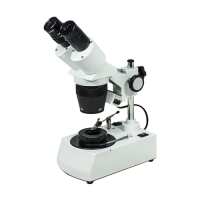
Quick OverviewFinite. Total Magnification: 20X/40X. 10X Eyepiece. Eye Tube Angle: 45°. Eyepiece Field of View:
Dia. 20mm. Illumination Type: II Dual Illuminated Light. Top Illumination: Oblique Top Light. Bottom Illumination: Bright/Dark Field. Input Voltage: AC 110V 60Hz. Input Voltage: AC 120V 60Hz. Suggested ApplicationsJewelry & Gemology , Appraisal, Gemology, Jewelry Repair, Stone SettingJM12060201 20X/40X Binocular Dual Power Darkfield Jewelry MicroscopeOptical System SpecificationsOptical SystemFiniteSystem Optical Magnification20X/40XTotal Magnification20X/40XStandard Eyepiece10X EyepieceSystem Field of View Dia. 5mm/ Dia. 10mmSystem Working Distance100mmBinocular Dual Power BodyBody Optical SystemFiniteBody Magnification2X/4XEye Tube Adjustment ModeSiedentopfEye Tube Angle45°Erect/Inverted ImageErect imageEye Tube Rotatable360° Degree RotatableInterpupillary Adjustment54-75mmEye Tube Inner Diameter Dia. 30.5mmEye Tube Diopter AdjustableLeft ±5°,
Right Not AdjustableEye Tube Fixing ModeLocking ScrewEyepiece TypeStandard EyepieceEyepiece Optical Magnification10XPlan EyepiecePlan EyepieceEyepiece Size for Eye Tube Dia. 30.5mmEyepiece Field of View Dia. 20mmPost StandVertical Post Height155mmVertical Post Diameter Dia. 14mmBase TypeIllumination BaseBase Dimensions190x115x55mmFocus ModeManualFocus Distance37mmCoarse Focus Distance per Rotation18mmIllumination TypeII Dual Illuminated LightTop IlluminationOblique Top LightTop Illumination TypeIncandescent LightBottom IlluminationBright/Dark FieldBottom Illumination TypeIncandescent LightInput VoltageAC 120V 60HzJewel TweezersJewel Tweezers TypeSteel Wire ClampJewel Tweezers Mounting Thread Size Dia. 3mmSurface TreatmentPolished ChromeMaterialMetalColorSilverNet Weight0.02kg (0.04lbs)Applied FieldFor JM1201, JM1206 Series MicroscopeDarkfield AttachmentDarkfield Condenser for Stereo MicroscopeDarkfield Attachment TypeDark FieldDarkfield Attachment Mount Size Dia. 95x5mmSurface TreatmentElectroplating BlackMaterialAluminumColorBlackNet Weight0.14kg (0.30lbs)Halogen Bulb12V 10W Halogen BulbBulb Rated Power10WBulb Rated VoltageDC 12VBulb ShapeOvalBulb Mounting ModeBi-PinMaterialGlassApplied FieldFor FS12 Series MicroscopePower SupplyOutput Power20WInput VoltageAC 110V 60HzOutput VoltageDC 12VPower Cord Connector TypeUSA 3 PinsPower Cable Length1.8mOther ParametersSurface TreatmentElectroplating BlackMaterialMetalColorBlackNet Weight2.70kg (5.95lbs)Dimensions190x140x310mm (7.480x5.512x12.205 in. )SeriesJM1206JM12060201
.tb_part
{
width: 100%;
border: solid 0px #ccc;
font-size:12px;
}
.tb_part tr
{
height: 30px;
line-height: 30px;
}
.tb_part td
{
white-space: nowrap;
}
.tb_part_1 td
{
white-space: wrap;
}
.normal{
border-left:solid 2px black;
border-right:solid 2px black;
}
.tb_part tr.normal td
{
border:solid 1px #ccc;
padding-right:5px;
}
.tb_part tr.normal:nth-child(2){
border-top:solid 2px black;
}
.tb_part tr.normal:first-child{
border:none;
}
.tb_part tr.normal:first-child td{
border:none;
font-weight:bold;
font-size:16px;
}
.end-border{
border-bottom:solid 2px black;
}
.tb_part tr.memo,.tb_part tr.memo td{
border:none;
}
Technical InfoInstructionsGemology/Jewelry MicroscopeClose ΛGemology/jewelry microscope is a microscope used to observe some of the invisible details on the surface and inside of jewels, gems, and various crystals when observing.
The surface features of jewellery, include scratches, breakage, color circles, etches, cleavage, cracks, split faces, polish, etc.; internal features, include the types of contents, the inclusions, growth lines, double crystal lines, textures, ribbons, and the doubling of the rear facet edge of the gemstone etc. For these features, it is very necessary to use gemology/jewelry microscope for observation and identification, and it has also an extremely important application in diamond grading, making it an essential instrument for gem identification.
For general jewellery observations, the defects and features after 10X magnification are considered the basis for economic evaluation. For jewelry microscope, stereo microscope is generally used, with a magnification of 10-80X or higher, and more detailed features can be seen. According to the characteristics of jewelry observation, jewelry microscope requires large depth of field, which can present the diamonds, jewelry interiors and their polyhedral structure, with true color reproduction effect.
Due to the different types of gemstones being observed, jewelry microscope needs to be matched with a variety of light sources and illumination forms. For lighting, usually full-color light is used, its continuous spectrum reflects the true color of the jewelry, and can achieve good observation effect for transparent, translucent and opaque object. The lighting methods of gemology microscope mainly include: Reflective illumination: use the reflector lamp above to observe opaque or translucent objects, as well as the surface features of the gem, such as the ports, cleavage surfaces, ribbons, and gemstone grinding etc. Transmitted illumination method: the transmitted light at the bottom is used to observe the internal features and defects of the gemstone. Usually, the bottom light is a diffuse reflection light source with dark field illumination, and there is a switch button, in the center of the bottom light, there is a black disc "light block", used to switch the light and dark field of view conversion of the bottom light.
Transmitted bright field illumination: the transmitted light can penetrate the interior of a gemstone. Against a bright background, details within the jewellery such as inclusions, textures, growth lines and ribbons can be seen clearly.
Transmitted light field dark illumination: after the bottom light is blocked by the "light block", it cannot directly enter the field of view of the objective lens, forming a diffuse reflection illuminating the gem around it, which is convenient for observing the inclusions, cracks and growth lines of the gemstone, and seeing the details that are not easy to find in bright field.
Spot light illumination method: adjust the aperture diaphragm (aperture) to a position close to the smallest suitable size, so that the bottom light is reduced into a dot shape, and the light penetrates from the bottom of the gemstone for illumination, so that the ribbon and the structural feature points of the gemstone are more prominent. Horizontal illumination method: use a sidelight to illuminate the gemstone horizontally from the side, so that the gem dot-like inclusions, textures, and bubbles etc. present striking and bright image under the vertical direction light illumination. Masking illumination method: use the bottom light bright field for illumination, insert an opaque light barrier in the field of view to increase the three-dimensional sense of the internal feature points in the black and white boundary of the light, which helps to observe the growth structure of the gem, such as the bending growth lines, double crystal lines, etc. Scattering illumination method: use the bottom light bright field for illumination, place a tissue or other translucent material on the light source to make the light softer, which helps to observe the color gamut and the ribbon, especially to observe the diffusion treatment effect of the gemstone. Polarized illumination method: under the bottom light source and the objective lens, a polarizer piece is added respectively to produce a polarizing effect, which can help to observe the interference effect, pleochroism and the optical characteristics of the gemstone.
For more precautions for use of gemology/jewelry microscopes, please refer to the Stereo Microscope on the BoliOptics website. FiniteClose ΛMicroscopes and components have two types of optical path design structures. One type is finite optical structural design, in which light passing through the objective lens is directed at the intermediate image plane (located in the front focal plane of the eyepiece) and converges at that point. The finite structure is an integrated design, with a compact structure, and it is a kind of economical microscope. Another type is infinite optical structural design, in which the light between the tube lens after passing the objective lens becomes "parallel light". Within this distance, various kinds of optical components necessary such as beam splitters or optical filters call be added, and at the same time, this kind of design has better imaging results. As the design is modular, it is also called modular microscope. The modular structure facilitates the addition of different imaging and lighting accessories in the middle of the system as required. The main components of infinite and finite, especially objective lens, are usually not interchangeable for use, and even if they can be imaged, the image quality will also have some defects.
The separative two-objective lens structure of the dual-light path of stereo microscope (SZ/FS microscope) is also known as Greenough. Parallel optical microscope uses a parallel structure (PZ microscope), which is different from the separative two-object lens structure, and because its objective lens is one and the same, it is therefore also known as the CMO common main objective. System Optical MagnificationClose ΛThe magnification of the objective lens refers to the lateral magnification, it is the ratio of the image to the real size after the original image is magnified by the instrument. This multiple refers to the length or width of the magnified object. System optical magnification is the product of the eyepiece and the objective lens (objective lens zoom set) of the optical imaging part within the system. Optical magnification = eyepiece multiple X objective lens/objective lens set
The maximum optical magnification of the microscope depends on the wavelength of the light to which the object is illuminated. The size of the object that can be observed must be greater than the wavelength of the light. Otherwise, the light cannot be reflected or transmitted, or recognized by the human eye. The shortest wavelength of ultraviolet light is 0.2 microns, so the resolution of the optical microscope in the visible range does not exceed 0.2 microns, or 200 nanometers. This size is converted to the magnification of the microscope, and it is the optical magnification of 2000X. Usually, the compound microscope can achieve 100X objective lens, the eyepiece is 20X, and the magnification can reach 2000X. If it is bigger, it will be called "invalid magnification", that is, the image is large, but the resolution is no longer increased, and no more details and information can be seen. Total MagnificationClose ΛTotal magnification is the magnification of the observed object finally obtained by the instrument. This magnification is often the product of the optical magnification and the electronic magnification. When it is only optically magnified, the total magnification will be the optical magnification.
Total magnification = optical magnification X electronic magnification Total magnification = (objective X photo eyepiece) X (display size / camera sensor target ) System Field of ViewClose ΛField of View, is also called FOV.
The field of view, or FOV, refers to the size of the object plane (i.e., the plane of the point of the observed object perpendicular to the optical axis), or of its conjugate plane (i.e., object to primary image distance), represented by a line value. System field of view is the size of the actual diameter of the image of the terminal display device of the instrument, such as the size of the image in the eyepiece or in the display.
Field of view number refers to the diameter of the field diaphragm of the objective lens, or the diameter of the image plane formed by the field diaphragm. Field of view number of objective lens = field of view number of eyepiece / (objective magnification / mechanical tube length)
Large field of view makes it easy to observe the full view and more range of the observed object, but the field of view (FOV) is inversely proportional to the magnification and inversely proportional to the resolution, that is, the larger the field of view, the smaller the magnification, and also the lower the resolution of the object to be observed. There are usually two ways to increase the field of view, one is to replace with an objective lens of a smaller multiple, or to replace with an eyepiece of a smaller multiple. System Working DistanceClose ΛWorking distance, also referred to as WD, is usually the vertical distance from the foremost surface end of the objective lens of the microscope to the surface of the observed object. When the working distance or WD is large, the space between the objective lens and the object to be observed is also large, which can facilitate operation and the use of corresponding lighting conditions.
In general, system working distance is the working distance of the objective lens. When some other equipment, such as a light source etc., is used below the objective lens, the working distance (i.e., space) will become smaller.
Working distance or WD is related to the design of the working distance of the objective lens. Generally speaking, the bigger the magnification of the objective lens, the smaller the working distance. Conversely, the smaller the magnification of the objective lens, the greater the working distance. When it is necessary to change the working distance requirement, it can be realized by changing the magnification of the objective lens. Binocular Dual Power BodyClose ΛBinocular dual power body refers to the main body of a stereo microscope with two objective lens magnifications. When performing magnification-shifting, two different magnifications can be obtained. When the body needs other different magnifications, it can also be solved by adding or changing the objective lens/auxiliary objective lens, or by changing the eyepieces of different magnifications. Dual power stereo microscope mostly uses 10X, 20X, 30X, 40X combinations of two kinds of magnifications. When the magnification is greater than 40X, the image is close to the plane effect, and the stereoscopic effect is relatively poor. Dual power stereo microscope has a simple structure, high reliability and low cost, it is a kind of stereo microscope that can satisfy stereo image applications. SiedentopfClose ΛFor siedentopf eyetube, when changing the interpupillary distance, it requires two hands pushing or pulling the two eyetubes left and right simultaneously, and the two eyepiece tubes or eyetubes will change their position at the same time.Eye Tube AngleClose ΛUsually the Microscope Eyetube is 45°, some is 30°, Tiltable Eyetube Angle design of a microscope is also known as the ergonomics microscope. 0-30° or 0-45° is an ergonomic design. When the mechanical tube length / focal length of the tube of the microscope is relatively big, the microscope is relatively high, and the user's height or the seat of the work desk is not suitable, long-term use of microscope may cause sitting discomfort. Eyepiece tube with variable angle can freely adjust the angle without lowering the head. Especially when it is close to 0 degree and the human eye is close to horizontal viewing, long-time or long-term use can avoid fatigue damage to the cervical vertebra.Erect/Inverted ImageClose ΛAfter imaging through a set of objective lenses, the object observed and the image seen by the human eye is inverted. When the observed object is manipulated, move the specimen or object, the image will move in the opposite direction in the field of view. Most of the biological microscopes are reversed-phase designs. When needing to operate works with accurate direction, it is necessary to design it into a forward microscope. Generally stereo microscopes and metallurgical microscopes are all of erect image design. When observing through the camera and display, the erect and inverted image can be changed by the orientation of the camera. 360° Degree RotatableClose ΛThe eyepiece of the microscope can have different viewing or observing directions. When the position of the microscope is uncomfortable, the direction of the eyepiece tube of the microscope can be adjusted, to facilitate observation and operation.
Placement method of different viewing angles of the microscope: General direction: the support column is behind the object to be observed Reverse direction: the support column is in front of the object to be observed Lateral direction: the support column is on the side of the object to be observed Rotating eyepiece tube, different microscopes may have different methods, for some, the direction is confirmed when installing the eyepiece tube of the microscope, for some, by rotating the body of the microscope, and for some, by rotating the support member on the support or holder of the microscope.Interpupillary AdjustmentClose ΛThe distance between the two pupils of the human eye is different. When the image of exit pupil of the two eyepieces of the microscope are not aligned with the entry pupil of the eye, the two eyes will see different images, which can cause discomfort. Adjust the distance between the two eyepieces, to accommodate or adapt to the pupil distance of the observer's eyes. The adjustment range is generally between 55-75mm. Eye Tube Diopter AdjustableClose ΛFor most people, their two eyes, the left and the right, have different vision; for the eyepiece tube, the eyepoint height of the eyepiece can be adjusted to compensate for the difference in vision between the two eyes, so that the imaging in the two eyes is clear and consistent.
The range of adjustment of the eyepiece tube is generally diopter plus or minus 5 degrees, and the maximum differential value between the two eyepieces can reach 10 degrees.
Monocular adjustable and binocular adjustable: some microscopes have one eyepiece tube adjustable, and some have two eyepiece tubes adjustable. First, adjust one eyepiece tube to the 0 degree position, adjust the microscope focusing knob, and find the clear image of this eyepiece (when the monocular adjustable is used, first adjust the focusing knob to make this eyepiece image clear), then adjust the image of another eyepiece tube (do not adjust the focusing knob again at this time), repeatedly adjust to find the clear position, then the two images are clear at the same time. For this particular user, do not adjust this device anymore in the future. As some microscopes do not have the vision adjustment mechanism for the eyepiece tube, the vision of the two eyes are adjusted through the eyepiece adjustable.
Eyepiece Optical MagnificationClose ΛEyepiece optical magnification is the visual magnification of the virtual image after initial imaging through the eyepiece. When the human eye observes through the eyepiece, the ratio of the tangent of the angle of view of the image and the tangent of the angle of view of the human eye when viewing or observing the object directly at the reference viewing distance is usually calculated according to 250 mm/focal length of eyepiece. The standard configuration of a general microscope is a 10X eyepiece. Usually, the magnification of the eyepiece of compound microscope is 5X, 8X, 10X, 12.5X, 16X, 20X. As stereo microscope has a low total magnification, its eyepiece magnification generally does not use 5X, but can achieve 25X, 30X and other much bigger magnification. Eyepiece Field of ViewClose ΛThe eyepiece field of view is the diameter of the field diaphragm of the eyepiece, or the diameter of the image plane of the field diaphragm imaged by the field diaphragm. The diameter of a large field of view can increase the viewing range, and see more detail in the field of view. However, if the field of view is too large, the spherical aberration and distortion around the eyepiece will increase, and the stray light around the field of view will affect the imaging effect. Post StandClose ΛPost stand generally has relatively tall post. When the focus is adjusted, the focusing mechanism can slide up and down the post, the microscope is thus placed in an approximately focused position, and then the focusing mechanism makes fine and accurate adjustment. This kind of stand can move quickly, and is suitable for viewing objects with a higher height and bigger volume. After the microscope is mounted, the microscope imaging center needs to be aligned with the center of the platen. The focusing mechanism button on the post must be tightened to lock the guard ring device, and the microscope should be prevented from loosening and shaking when working. When it is necessary to adjust the height, hold the microscope and the focusing mechanism with one hand, then release the knob, adjust it to the proper position, lock the knob, then top the guard ring to the lower position of the focusing mechanism, and lock it tight. In particular, avoid accidental dropping of the microscope due to gravity, thereby damaging the microscope and the objects below. Illumination BaseClose ΛIllumination base is a modular light source component, suitable for microscope stand base that has no light source of itself, and it is usually dedicated components supporting some stands. Illumination base typically includes at least one bottom lighting, and there are also illumination base that includes the circuit portion of the upper light source. Jewel TweezersClose ΛJewel tweezers are clips used to clamp jewels or diamonds. The spring clip at the front end can be freely opened and closed to clamp the jewel, the rear end is often fixed on the base, and can be freely rotated and stretched to observe the different positions and angles of the jewel. The commonly used wire gemstone clamps, have moderate steel property, strong clamping, and can reduce the light shielding of the gemstone waist, allowing comprehensive viewing of the gemstone in all directionsDark FieldClose ΛBright field illumination is that direct light shining on the object to be observed and the background enters directly into the objective lens, producing a background of a bright image; the dark field corresponds to the bright field, so that the light is obliquely irradiated onto the surface of the sample, and the direct light is not allowed to pass through the aperture of the objective lens, so as to be able to observe object details in a black background.
The light source adopts parallel light, and an annular visor is placed at an appropriate position on the incident light path to cover the central portion of the light. The surrounding light passing through the annular visor is a hollow cylindrical beam and is incident on the vertical illuminator. The vertical illuminator of the dark field illumination is a circular reflector that reflects the cylindrical beam upwards and projects it onto the reflector of the condenser, so that the reflected light is concentrated on the surface of the sample. Since the reflected light is highly tilted, the light cannot enter the objective lens, therefore the field of view is dark. Only in the concave place of the observed object can light enter the objective lens, and the bright white image is reflected in the dark field of view, called "dark field of view " (dark field) illumination. In most cases, the bright portion of the black-and-white image obtained by dark field illumination is considered to be the black portion of the black-and-white image obtained by bright field illumination. Dark field illumination improves the contrast of the image, making the color of the image natural and uniform. The angle of the incident beam is extremely large, which increases the effective numerical aperture of the objective lens, and the ability to identify details is higher than that of bright field illumination. Dark field illumination must ensure that the system is clean, as dust particles can form bright spots in the dark field, which can affect the results of observation. PackagingClose ΛAfter unpacking, carefully inspect the various random accessories and parts in the package to avoid omissions. In order to save space and ensure safety of components, some components will be placed outside the inner packaging box, so be careful of their inspection. For special packaging, it is generally after opening the box, all packaging boxes, protective foam, plastic bags should be kept for a period of time. If there is a problem during the return period, you can return or exchange the original. After the return period (usually 10-30 days, according to the manufacturer’s Instruction of Terms of Service), these packaging boxes may be disposed of if there is no problem.
About Boli Optics:
Boli Optics Microscope Supplier sells professional microscopes, microscope accessories, and magnifying lamps. We offer parts and accessories compatible with Leica, Olympus, Nikon, and Zeiss, and more. We supply research laboratories, medical centers, universities, industrial manufactures, factories, and more. Our engineers and technicians provide technical support and design & manufacture custom microscope products for your applications. Our products are manufactured under ISO 9001 quality control standards. We also provide OEM service. Since 1994, our talented team has been working in the optics industry and serving our customers whole-heartedly. Based in Southern California, we offer fast, same day shipping from our local warehouses.
Visit Product PageEmail:
sales@bolioptics.com
Location:
8762 Lanyard Court,
Rancho Cucamonga
California
91730