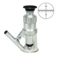
Instruction ManualPM4201 Portable Measurement Microscope Instruction Manual-English.docQuick OverviewFinite. Total Magnification: 60X. 10X Eyepiece. 6X Achromatic Objective. Eyepiece Field of View:
Dia. 12mm. Illumination Type: LED Reflection Light. Top Illumination: Oblique Top Light. Suggested ApplicationsEnvironmental , Field Research, Forensics , Counterfeit Currency DetectionPM42010312 60X Portable Measurement MicroscopeOptical System SpecificationsOptical SystemFiniteMechanical Tube Length160mmSystem Optical Magnification60XExpandable System Optical Magnification (Optional Parts Required)20X/40X/60X/100XTotal Magnification60XStandard Eyepiece10X EyepieceStandard Objective6X Achromatic ObjectiveEyepieceEyepiece TypeStandard EyepieceEyepiece Optical Magnification10XPlan EyepiecePlan EyepieceEyepiece Size for Eye Tube Dia. 23mmEyepiece Field of View Dia. 12mmEyepiece Size for Reticle Dia. 16mmReticle TypeX-Axis Crosshair ScaleReticle Dimensions Dia. 16x0.5mmObjectiveObjective Optical SystemFiniteObjective Optical Magnification6XObjective Parfocal Distance45mmObjective Working Distance13.5mmNumerical Aperture (N.A.)N.A. 0.25Microscope StandStand Height200mmFocus ModeManualFocus Distance42mmCoarse Focus Distance per Rotation21mmFocusing Knob Tightness AdjustableTightness Not AdjustableMicroscope IlluminatorIllumination TypeLED Reflection LightTop IlluminationOblique Top LightTop Illumination TypeLEDOther ParametersSurface TreatmentNatural OxidationMaterialAluminumColorWhiteNet Weight0.60kg (1.32lbs)Dimensions Dia. 63x180mm ( Dia. 2.480x7.087 in. )
.tb_part
{
width: 100%;
border: solid 0px #ccc;
font-size:12px;
}
.tb_part tr
{
height: 30px;
line-height: 30px;
}
.tb_part td
{
white-space: nowrap;
}
.tb_part_1 td
{
white-space: wrap;
}
.normal{
border-left:solid 2px black;
border-right:solid 2px black;
}
.tb_part tr.normal td
{
border:solid 1px #ccc;
padding-right:5px;
}
.tb_part tr.normal:nth-child(2){
border-top:solid 2px black;
}
.tb_part tr.normal:first-child{
border:none;
}
.tb_part tr.normal:first-child td{
border:none;
font-weight:bold;
font-size:16px;
}
.end-border{
border-bottom:solid 2px black;
}
.tb_part tr.memo,.tb_part tr.memo td{
border:none;
}
Technical InfoInstructionsPortable MicroscopeClose ΛPortable microscope is the general term for microscopes that are simple in design, easy to carry and convenient for field observation. It usually does not refer to a certain kind of microscope.
Portable microscope is simple in design, but also like a microscope, there is at least one eyepiece and one objective lens, or is imaged by a camera, and has a stand and focusing device. Most of them can also be connected to a camera or an eyepiece camera, and then connect the monitor or store digital images. Portable microscope generally adopts hand-held operation, and has simple configuration, fixed working distance, convenient for quick observation. Generally, portable microscope has a light source with a battery, which is convenient to carry to work place and field work, and is suitable for application of various industries and scenarios. FiniteClose ΛMicroscopes and components have two types of optical path design structures. One type is finite optical structural design, in which light passing through the objective lens is directed at the intermediate image plane (located in the front focal plane of the eyepiece) and converges at that point. The finite structure is an integrated design, with a compact structure, and it is a kind of economical microscope. Another type is infinite optical structural design, in which the light between the tube lens after passing the objective lens becomes "parallel light". Within this distance, various kinds of optical components necessary such as beam splitters or optical filters call be added, and at the same time, this kind of design has better imaging results. As the design is modular, it is also called modular microscope. The modular structure facilitates the addition of different imaging and lighting accessories in the middle of the system as required. The main components of infinite and finite, especially objective lens, are usually not interchangeable for use, and even if they can be imaged, the image quality will also have some defects.
The separative two-objective lens structure of the dual-light path of stereo microscope (SZ/FS microscope) is also known as Greenough. Parallel optical microscope uses a parallel structure (PZ microscope), which is different from the separative two-object lens structure, and because its objective lens is one and the same, it is therefore also known as the CMO common main objective. Mechanical Tube LengthClose ΛFor objective lens design of finite microscope, its mechanical tube length is the distance from the objective nosepiece shoulder of the objective lens to the eyepiece seat in the tubes, that is, the eyepiece shoulder.
There are two standards in the traditional microscope structure, namely, DIN and JIS. DIN (Deutsches Institute fur Normung) is a popular international standard for microscopes, using 195mm standard conjugate distance (also known as object to primary image distance, 36mm objective lens parfocal distance, and 146.5mm optical tube length.
JIS (Japanese Industrial Standard) is a standard adopted by some Japanese manufacturers, using 160mm standard conjugate distance (also known as object to primary image distance), 45mm objective lens parfocal distance), and 150mm optical tube length.
Using the same microscope standard design, the objective lenses can be used interchangeably. System Optical MagnificationClose ΛThe magnification of the objective lens refers to the lateral magnification, it is the ratio of the image to the real size after the original image is magnified by the instrument. This multiple refers to the length or width of the magnified object. System optical magnification is the product of the eyepiece and the objective lens (objective lens zoom set) of the optical imaging part within the system. Optical magnification = eyepiece multiple X objective lens/objective lens set
The maximum optical magnification of the microscope depends on the wavelength of the light to which the object is illuminated. The size of the object that can be observed must be greater than the wavelength of the light. Otherwise, the light cannot be reflected or transmitted, or recognized by the human eye. The shortest wavelength of ultraviolet light is 0.2 microns, so the resolution of the optical microscope in the visible range does not exceed 0.2 microns, or 200 nanometers. This size is converted to the magnification of the microscope, and it is the optical magnification of 2000X. Usually, the compound microscope can achieve 100X objective lens, the eyepiece is 20X, and the magnification can reach 2000X. If it is bigger, it will be called "invalid magnification", that is, the image is large, but the resolution is no longer increased, and no more details and information can be seen. Total MagnificationClose ΛTotal magnification is the magnification of the observed object finally obtained by the instrument. This magnification is often the product of the optical magnification and the electronic magnification. When it is only optically magnified, the total magnification will be the optical magnification.
Total magnification = optical magnification X electronic magnification Total magnification = (objective X photo eyepiece) X (display size / camera sensor target ) Eyepiece Optical MagnificationClose ΛEyepiece optical magnification is the visual magnification of the virtual image after initial imaging through the eyepiece. When the human eye observes through the eyepiece, the ratio of the tangent of the angle of view of the image and the tangent of the angle of view of the human eye when viewing or observing the object directly at the reference viewing distance is usually calculated according to 250 mm/focal length of eyepiece. The standard configuration of a general microscope is a 10X eyepiece. Usually, the magnification of the eyepiece of compound microscope is 5X, 8X, 10X, 12.5X, 16X, 20X. As stereo microscope has a low total magnification, its eyepiece magnification generally does not use 5X, but can achieve 25X, 30X and other much bigger magnification. Eyepiece Field of ViewClose ΛThe eyepiece field of view is the diameter of the field diaphragm of the eyepiece, or the diameter of the image plane of the field diaphragm imaged by the field diaphragm. The diameter of a large field of view can increase the viewing range, and see more detail in the field of view. However, if the field of view is too large, the spherical aberration and distortion around the eyepiece will increase, and the stray light around the field of view will affect the imaging effect. Objective Optical MagnificationClose ΛThe finite objective is the lateral magnification of the primary image formed by the objective at a prescribed distance.
Infinite objective is the lateral magnification of the real image produced by the combination of the objective and the tube lens.
Infinite objective magnification = tube lens focal length (mm) / objective focal length (mm)
Lateral magnification of the image, that is, the ratio of the size of the image to the size of the object. The larger the magnification of the objective, the higher the resolution, the smaller the corresponding field of view, and the shorter the working distance. Objective Parfocal DistanceClose ΛObjective parfocal distance refers to the imaging distance between the objective shoulder and the uncovered object surface (referred to as the “object distance). It conforms to the microscope design, usually 45mm.
The objective of different magnifications of the compound microscope has different lengths; when the distance between the objective shoulder and the object distance is the same, the focal length may not be adjusted when converting to objectives of different magnifications.
Objective Working DistanceClose ΛThe objective working distance is the vertical distance from the foremost surface end of the objective of the microscope to the object surface to be observed. Generally, the greater the magnification, the higher the resolution of the objective, and the smaller the working distance, the smaller the field of view. Conversely, the smaller the magnification, the lower the resolution of the objective, and the greater the working distance, and greater the field of view.
High-magnification objectives (such as 80X and 100X objectives) have a very short working distance. Be very careful when focusing for observation. Generally, it is after the objective is in position, the axial limit protection is locked, then the objective is moved away from the direction of the observed object.
The relatively greater working distance leaves a relatively large space between the objective and the object to be observed. It is suitable for under microscope operation, and it is also easier to use more illumination methods. The defect is that it may reduce the numerical aperture of the objective, thereby reducing the resolution.
Numerical Aperture (N.A.)Close ΛNumerical aperture, N.A. for short, is the product of the sinusoidal function value of the opening or solid angle of the beam reflected or refracted from the object into the mouth of the objective and the refractive index of the medium between the front lens of the objective and the object. Simply speaking, it is the magnitude of the luminous flux that can be brought in to the mouth of the objective adapter, the closer the objective to the specimen for observation, the greater the solid angle of the beam entering the mouth of the objective adapter, the greater the N.A. value, and the higher the resolution of the objective. When the mouth of the objective adapter is unchanged and the working distance between the objective and the specimen is constant, the refractive index of the medium will be of certain meaning. For example, the refractive index of air is 1, water is 1.33, and cedar oil is 1.515, therefore, when using an aqueous medium or cedar oil, a greater N.A. value can be obtained, thereby improving the resolution of the objective.
Formula is N.A. = refractive index of the medium X sin solid angle of the beam of the object entering the front lens frame of the objective/ 2
Numerical aperture of the objective. Usually, there is a calculation method for the magnification of the microscope. That is, the magnification of the microscope cannot exceed 1000X of the objective. For example, the numerical aperture of a 100X objective is 1.25, when using a 10X eyepiece, the total magnification is 1000X, far below 1.25 X 1000 = 1250X, then the image seen in the eyepiece is relatively clear; if a 20X eyepiece is used, the total magnification will reach 2000X, much higher than 1250X, then eventhoughthe image actually seen by the 20X eyepiece is relatively large, the effect will be relatively poor. Focusing Knob Tightness AdjustableClose ΛDifferent microscope bodies, different human operations, and different requirements for observation and operation, all require adjustment of the pre-tightening force of the stand that support microscope body. Facing the stand just right, use both hands to reverse the force to adjust the tightness. (face the knob of one side just right, clockwise is to tighten, counterclockwise is to loosen) In general, after long-time use, the knob will be loose, and adjustment is necessary.
PackagingClose ΛAfter unpacking, carefully inspect the various random accessories and parts in the package to avoid omissions. In order to save space and ensure safety of components, some components will be placed outside the inner packaging box, so be careful of their inspection. For special packaging, it is generally after opening the box, all packaging boxes, protective foam, plastic bags should be kept for a period of time. If there is a problem during the return period, you can return or exchange the original. After the return period (usually 10-30 days, according to the manufacturer’s Instruction of Terms of Service), these packaging boxes may be disposed of if there is no problem.
Optical Data Microscope Optical Data SheetP/NObjectiveObjective Working DistanceEyepiecePM42010312 (10X Dia. 12mm)MagnificationField of View(mm)PM420103126X13.5mm60X2mm1. Magnification=Objective Optical Magnification * Body Magnification * Eyepiece Optical Magnification2. Field of View=Eyepiece Field of View /Objective Optical Magnification*Body Magnification3. The Darker background items are Standard items, the white background items are
optional items.
About Boli Optics:
Boli Optics Microscope Supplier sells professional microscopes, microscope accessories, and magnifying lamps. We offer parts and accessories compatible with Leica, Olympus, Nikon, and Zeiss, and more. We supply research laboratories, medical centers, universities, industrial manufactures, factories, and more. Our engineers and technicians provide technical support and design & manufacture custom microscope products for your applications. Our products are manufactured under ISO 9001 quality control standards. We also provide OEM service. Since 1994, our talented team has been working in the optics industry and serving our customers whole-heartedly. Based in Southern California, we offer fast, same day shipping from our local warehouses.
Visit Product PageEmail:
sales@bolioptics.com
Location:
8762 Lanyard Court,
Rancho Cucamonga
California
91730